Introduction
A stereo microscope (also called a dissecting microscope) is a specialized optical instrument designed to provide a three-dimensional view of specimens at low to moderate magnification. Unlike compound microscopes that produce a single optical path and high magnification, stereo microscopes use two separate optical channels to deliver slightly different views to each eye, creating depth perception and spatial context. This makes them indispensable when performing tasks that require hand-eye coordination, such as dissection, assembly of small parts, quality inspection, and examining biological or geological samples. Modern stereo microscopes come with a range of illumination options, zoom or fixed magnification heads, ergonomic stands, and digital camera attachments that support documentation and collaboration. For anyone selecting or using a stereo microscope, understanding optical design, illumination choices, and practical handling will maximize performance and protect both instrument and specimens. The rest of this article breaks down core components, how they work, typical uses across industries, and smart buying and maintenance tips all written to help readers make confident, well-informed decisions.
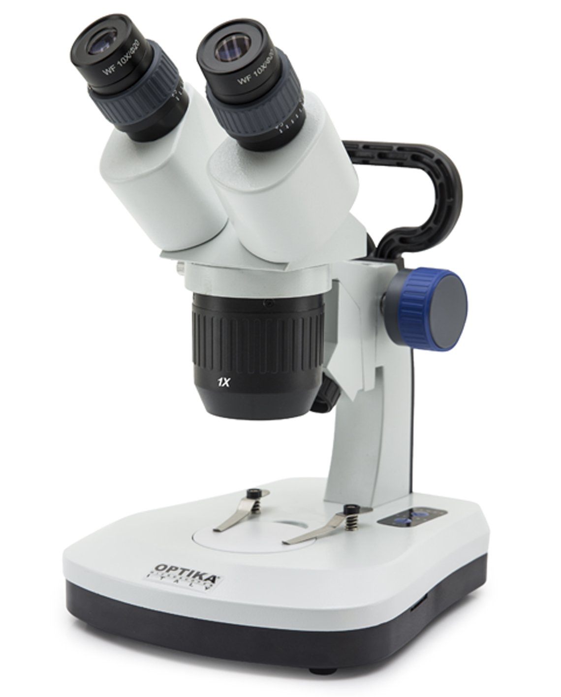
What is a stereo microscope and how does it differ from other microscopes?
A stereo microscope is engineered to provide a low-power, wide-field, three-dimensional image by using two separate optical paths one for each eye with independent objectives or a dual-channel optical system. This binocular design gives depth perception, making it easy to judge height, texture, and spatial relationships at the specimen surface. Typical magnifications range from about 5× to 80× (often achieved via a zoom head), which is ideal for macroscopic tasks where detail plus spatial context matter. In contrast, compound microscopes use a single optical path and much higher magnification to resolve cellular or subcellular structures on thin sections or slides; they usually produce flat images and require transmitted light through the sample. Stereo microscopes often use reflected (incident) light from above for opaque samples, though many models incorporate transmitted light bases for semi-transparent specimens. The result is a more intuitive, real-world view useful for manipulation, inspection, and documentation tasks for which compound microscopes are not well suited.
Key components and optical principles that determine performance
A stereo microscope’s performance depends on its optical head, objectives, eyepieces, zoom mechanism, illumination, and stand. The optical head houses the binocular tubes and prisms that deliver two offset images; prism quality affects image brightness and color fidelity. Eyepieces (commonly 10×) pair with objective optics usually a fixed objective plus a zoom range to produce final magnification. Zoom heads provide continuous magnification adjustment and are described by their zoom ratio (e.g., 6.7:1), which influences how smoothly you can frame a subject. Illumination is equally important: incident LED rings or spot lights illuminate opaque objects from above, while built-in transmitted light bases let you view thin or semi-transparent samples. Stands range from simple hinges to heavy-duty boom stands that allow large part inspection. Additional features that influence practical performance include long working distance objectives for easy tool access, parfocal zoom for minimal refocusing during magnification changes, and the option to attach digital cameras for imaging and measurement. Understanding each component helps match a microscope to its intended use.
Practical applications
Stereo microscopes are widely used across many disciplines because their 3D view and comfortable working distance enable detailed manual work and inspection. In biology and education, they’re standard for dissections, specimen sorting, and teaching students can observe structures without preparing slides. In electronics and manufacturing, technicians use stereo microscopes for soldering, circuit board inspection, and microassembly because the view allows accurate tool placement and defect identification. In gemology and watchmaking, fine surface detail and depth cues are essential for appraisal and repair. Conservationists and archaeologists rely on gentle, magnified manipulation during restoration, while entomologists and botanists use stereo microscopes to study morphology without destroying specimens. Medical device manufacturers, forensic labs, and quality control departments also depend on stereo imaging for routine inspection and documentation. The combination of hands-on capability, depth perception, and adaptability (illumination and documentation options) makes the stereo microscope a practical choice wherever three-dimensional detail and manipulation intersect.
Choosing the right stereo microscope and maintaining it for longevity
Selecting the right stereo microscope starts with clarifying tasks: do you need wide field low magnification for assembly, or fine detail for inspection? Choose an appropriate zoom range and eyepiece magnification to reach required working magnifications without compromising field of view. Prioritize long working distance optics if you’ll use tools under the objective, and consider a boom or articulated stand for larger samples. Illumination matters: incident LED rings or fiber optic lights help with textured surfaces, while adjustable transmitted light is useful for thin specimens. If documentation is required, ensure camera compatibility or an integrated imaging head. For longevity and consistent performance, store the microscope covered in a dry, dust-free area; clean optical surfaces only with recommended lens tissue and solvent; avoid touching lenses with fingers; and have mechanical parts serviced if zoom action becomes stiff. Regularly check illumination LEDs and replace batteries or power supplies as needed. Following manufacturer maintenance guidelines preserves optical alignment and ensures reliable, long-term operation.
Conclusion
Stereo microscopes provide an approachable, highly practical bridge between unaided human vision and precise magnified observation, offering depth, comfort, and versatility. Their unique binocular design and range of illumination and stand options make them indispensable across education, biology, electronics, conservation, and quality control. Choosing the right model means aligning optical features zoom ratio, working distance, illumination type, and camera compatibility with the tasks you perform. Responsible maintenance and proper handling sustain performance and protect both instrument and specimens. Whether you’re a student, technician, researcher, or hobbyist, understanding these core principles will help you select, use, and care for a stereo microscope with confidence.
Frequently Asked Questions
Q: What magnification range do stereo microscopes typically offer?
A: Commonly from about 5× to 80×, often via a zoom head combined with eyepieces.
Q: Can I take photos with a stereo microscope?
A: Yes—many models accept C-mount cameras or have integrated camera ports for documentation.
Q: Which light type is best for inspecting circuit boards?
A: Incident (reflected) LED lighting and adjustable spot illumination work best for opaque electronic components.
Q: Do stereo microscopes need special cleaning?
A: Clean optics with lens tissue and recommended solvent; avoid harsh rubbing and cover when not in use.
Q: Is a stereo microscope good for teaching?
A: Absolutely its 3D view and ease of use make it ideal for labs, classrooms, and demonstrations.
Leave a Reply